

17th October 2026
How Does Digital Imaging Help Detect Mouth Cancer Early?
Every routine check-up includes a soft tissue examination, and for many patients it is the only time anyone looks closely inside their mouth.
So how does digital imaging help detect mouth cancer early? Well, first and foremost, it gives your clinicians a reliable way to record what they see, track changes between visits and share clear images with patients and specialists. Good surgery design then makes the examination itself easier to do well. With Mouth Cancer Action Month arriving in November, now is a sensible time to check whether your digital setup and surgery layout are pulling their weight.
The Overview:
- Mouth cancer diagnoses in England have reached a record high.
- A careful visual and tactile soft tissue exam remains the foundation of screening in general practice.
- Intraoral cameras let clinicians photograph, store and compare areas of concern across appointments.
- Lighting, chair access and screen placement decide how thorough an exam can be within a normal slot.
- Imaging upgrades and layout changes work best when they are planned together.
Planning An Imaging Upgrade or Surgery Refit?
The right equipment and layout make a thorough soft tissue check a natural part of every appointment. Explore our digital imaging range or get in touch to talk it through or to arrange a visit to our Hampshire showroom.
Why Is Early Detection Important for Mouth Cancer?
The Oral Health Foundation reported in November 2025 that 9,293 people in England were diagnosed with mouth cancer in a single year, a 37% rise over the past decade, with 2,970 deaths. The same charity found that almost a quarter of people don’t know cancer can develop in and around the mouth, and only around one in five recognise the most common signs.
Timing makes a huge difference to outcomes.
NHS Lanarkshire notes that around 10,000 people are diagnosed across the UK each year and that catching the disease early can lift survival from around 50% to 90%. Public Health Wales adds that the tongue and oropharynx remain the most common sites and that five-year survival stays below 50% for people diagnosed at an advanced stage.
With public awareness this low, the dental team is often the first to notice a change. That puts real weight on how well your surgery supports the examination.
How Dental Digital Imaging Supports Earlier Detection of Mouth Cancer
What dental imaging does is support the clinical exam. It does not diagnose mouth cancer, and suspicious lesions will still need specialist assessment and biopsy from a clinician. But the right equipment does help to record and communicate more clearly.
Intraoral Cameras for Documenting and Monitoring Lesions
A written note describing a small white patch on the side of the tongue is useful, but a clear photograph of the same patch is a much greater help. This is especially true when a patient returns for review and the clinician needs to judge whether anything has changed.
Intraoral images give your team a consistent visual record over time. When a referral is needed, attaching good images through the standard urgent referral route helps the receiving specialist understand the concern.
When photographs sit in the same imaging software as your radiographs and scans, everything stays in one patient record and is quick to find. Images also help patients. Seeing an area on screen makes it easier to understand why a review appointment or referral matters, which can encourage attendance.
Panoramic and CBCT Imaging for the Wider Picture
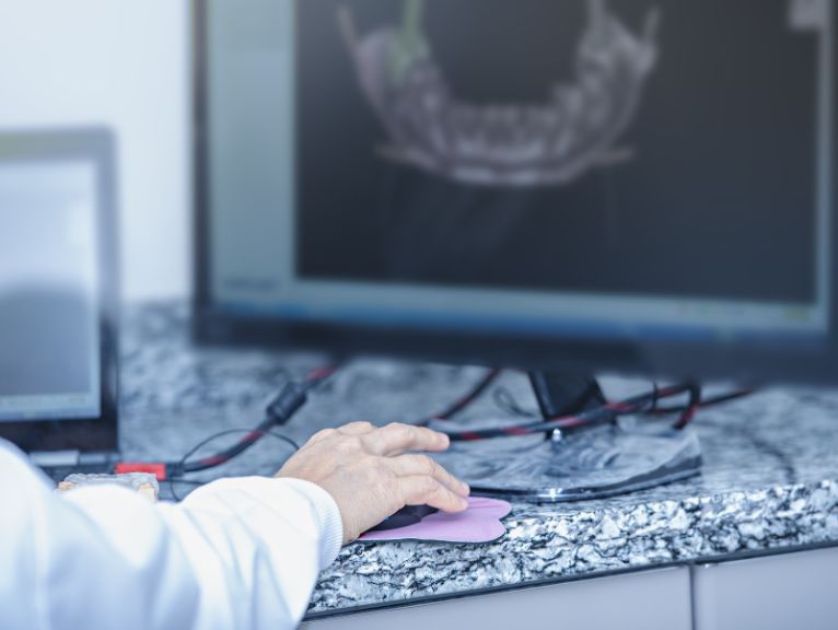
Panoramic radiographs and CBCT scans show bone and wider anatomy that a visual exam cannot reach. Systems like the Planmeca ProMax 3D give clinicians detailed views of the jaws when a clinical finding or a specialist request calls for it.
Neither shows early changes in the soft tissue lining of the mouth, which is why they have to sit alongside the soft tissue exam.
Dental Surgery Design for Thorough Oral Cancer Checks
A complete soft tissue exam covers the lips, cheeks, gums, palate, floor of the mouth, all surfaces of the tongue and the neck. Your surgery layout decides how comfortably that can happen.
Light Where the Clinician Needs It
Strong, even task lighting with accurate colour rendering helps dentists pick up the micro differences in tissue colour and texture. Ambient lighting matters too, so the operating light isn’t working against shadows and glare.
Chair Positioning and Ergonomic Access
Seeing the floor of the mouth and the underside of the tongue means positioning the patient and the clinician well. Treatment centres with slim backrests and flexible headrests let clinicians get close without straining, and supportive stools help them hold a good working posture through a full list.
Screens Patients Can See
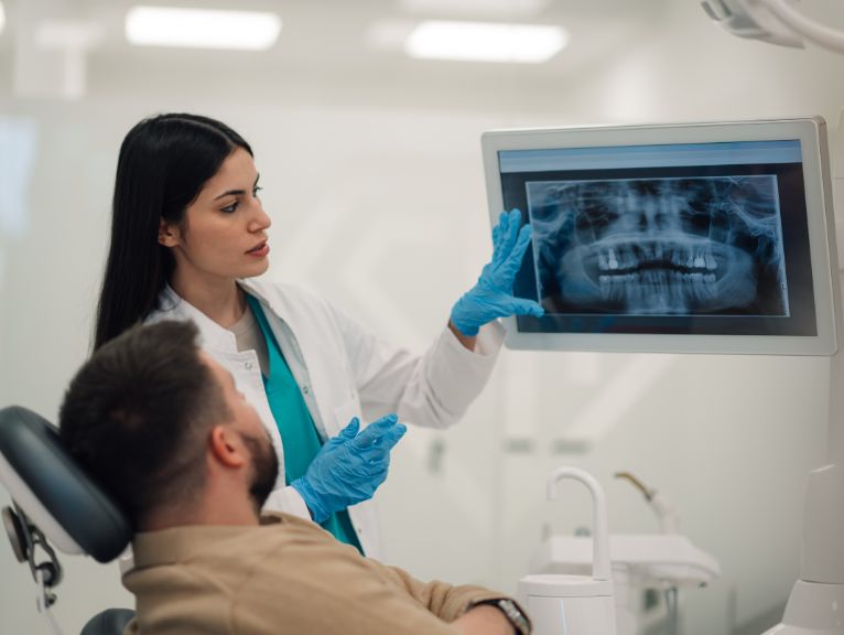
A chairside monitor placed within the patient’s line of sight lets the dentist show images clearly and explain what they are monitoring.
A Workflow That Fits the Appointment Slot
When the camera sits within reach on the delivery unit and images save straight to the patient record, documenting a concern takes minutes. Planned storage, cable routes and data points make this possible, and regular servicing through Curran Connected keeps cameras, lights and X-ray units ready for every patient.

Want to Strengthen Mouth Cancer Screening in Your Dental Practice?
Curran Dental has spent over 20 years designing, building and equipping dental practices across the UK. We supply digital imaging from manufacturers including Dürr Dental, Planmeca, and Acteon and our in-house design team plans surgeries around how your clinicians actually work, from lighting and chair position to screen placement and cable routes.
If you are reviewing your imaging or planning a refit ahead of Mouth Cancer Action Month, contact our team or explore our digital solutions to see what would suit your practice.
FAQs
How does digital imaging help detect mouth cancer early?
Digital imaging helps by recording what clinicians see during the soft tissue exam so changes can be tracked over time. Intraoral photographs make review appointments more objective and give specialists clear information when a referral is made. Imaging supports the clinical exam and specialist diagnosis rather than replacing them.
What role does an intraoral camera play in oral cancer screening?
An intraoral camera documents areas of concern so they can be monitored and compared at future visits. It also helps clinicians show patients what they have seen and share images with specialists. Diagnosis still depends on specialist assessment and biopsy.
What dental equipment helps with mouth cancer screening?
The most useful dental equipment for mouth cancer screening includes good operating and ambient lighting, an ergonomic treatment centre, an intraoral camera and a chairside screen patients can see. Imaging software that stores photographs with radiographs keeps the record in one place. Panoramic and CBCT imaging add information about bone and wider anatomy.
When is Mouth Cancer Action Month?
Mouth Cancer Action Month runs every November and is led by the Oral Health Foundation. It raises public awareness of the signs of mouth cancer and the value of regular dental check-ups. Many practices use it as a prompt to review their own screening routines.